Neurodegeneración
Aug 20, 2026
Sex differences in innate immune cell responses: Impact on cancer immunity and response to therapy
Sex-Dimorphic Innate Immune Responses in Neuroinflammation and Neurodegeneration: Molecular Pathways and Precision Biomarkers
Abstract
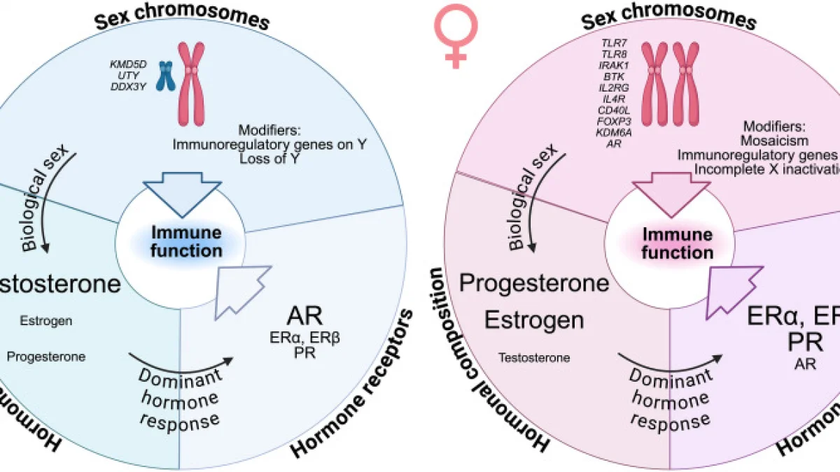
Innate immune cells, including resident microglia and infiltrating monocyte-derived macrophages, exhibit pronounced sex-dimorphic phenotypic shifts that fundamentally shape the pathogenesis and progression of neurodegenerative disorders. Biological sex dictates pattern recognition receptor responsiveness, toll-like receptor (TLR) signaling kinetics, and the activation thresholds of the NLRP3 inflammasome within the central nervous system. X-chromosome gene dosage effects—driven by incomplete X-chromosome inactivation of genes such as *TLR7*, *TLR8*, and *KDM6A*—interact synergistically with gonadal steroids (17β-estradiol and testosterone) to modulate neuroinflammatory cascades, lysosomal clearance capacity, and mitochondrial reactive oxygen species production. In females, heightened baseline microglial reactivity accelerates neuroinflammatory responses during neurodegenerative proteinopathy, whereas male innate myeloid cells demonstrate altered phagocytic efficiency and compromised tissue repair kinetics. Unraveling these sex-specific molecular mechanisms is imperative for establishing precision immunotherapies. Integrating sex-and-gender-based analysis (SGBA) into neuroimmunology provides a transformative framework for identifying sexually dimorphic biomarkers and refining therapeutic stratification in neurodegenerative pathology.
Introduction & Cellular Mechanisms
Innate immunity within the central nervous system (CNS) operates through a delicate equilibrium between homeostatic surveillance and inflammatory execution. Microglia, the primary parenchymal innate immune cells, together with perivascular macrophages and recruited circulating monocytes, serve as the first line of defense against pathological protein aggregates (e.g., amyloid-beta oligomers, hyperphosphorylated tau, and α-synuclein). However, persistent activation of these myeloid populations triggers toxic neuroinflammatory cascades that drive progressive neuronal loss.
At the molecular level, pathological ligands engage Pattern Recognition Receptors (PRRs), predominantly Toll-Like Receptors (TLR2, TLR4, TLR7/8) and C-type lectin receptors. Ligation of these receptors recruits the adaptor protein MyD88, initiating phosphorylation cascades involving the IκB kinase (IKK) complex. This sequence induces the nuclear translocation of the nuclear factor kappa B (NF-κB) p50/p65 heterodimer and the activation of Mitogen-Activated Protein Kinase (MAPK) pathways, upregulating pro-inflammatory cytokines such as interleukin-1 beta (IL-1β), interleukin-6 (IL-6), and tumor necrosis factor-alpha (TNF-α).
Concurrent with PRR priming, intracellular danger signals induce assembly of the NLRP3 inflammasome complex via ASC (apoptosis-associated speck-like protein containing a CARD) oligomerization and Caspase-1 activation. Caspase-1 cleaves pro-IL-1β and pro-IL-18 into their active secreted forms while executing gasdermin D-mediated pyroptosis.
```
[Pathological Protein Aggregates / DAMPs]
│
▼
[TLR2 / TLR4 / TLR7/8 Receptors]
│
(MyD88 Adaptor Recruitment)
│
▼
[IKK Phosphorylation Complex]
│
▼
[NF-κB (p50/p65) Translocation to Nucleus]
│
┌──────────────┴──────────────┐
▼ ▼
[Pro-inflammatory Cytokines] [NLRP3 Inflammasome Assembly]
(IL-1β, IL-6, TNF-α) │
▼
[Caspase-1 Activation]
│
▼
[Gasdermin D Pyroptosis & IL-1β/18 Secretion]
```
At the organellar level, neurodegenerative stimuli impair the autophagy-lysosome pathway (ALP) and mitochondrial dynamics. Microglial phagocytosis of proteopathic seeds requires functional lysosomal acidification and LC3-associated phagocytosis (LAP). Failure of lysosomal degradation leads to cytosolic leakage of cathepsins and continuous inflammasome propagation. Simultaneously, microglial mitochondrial dysfunction accelerates electron transport chain disruption, boosting mitochondrial reactive oxygen species (mtROS) generation. mtROS directly oxidizes mitochondrial DNA (mtDNA), which escapes into the cytosol to activate the cyclic GMP-AMP synthase (cGAS)-stimulator of interferon genes (STING) pathway, establishing a chronic, self-sustaining neuroinflammatory cycle.
Sex-Dimorphic Regulation & Translational Implications
Biological sex exerts profound control over the epigenome, transcriptome, and functional states of innate immune cells in the CNS. The dimorphism is driven by two distinct yet interacting mechanisms: genetic dosage from sex chromosomes and differential modulation by gonadal sex steroids.
1. Epigenetic and Genetic Architecture of Sex Dimorphism
The X chromosome encodes a disproportionate number of genes critical to immune function and chromatin remodeling. Although one X chromosome undergoes transcriptional silencing in mammalian females, approximately 15–20% of X-linked genes escape X-chromosome inactivation (XCI), leading to bi-allelic expression and heightened gene dosage in XX cells.
2. Hormonal Fine-Tuning of Neuroinflammatory Signaling
Circulating gonadal steroids cross the blood-brain barrier to bind nuclear and membrane-bound steroid receptors expressed on microglial and monocyte surfaces:
3. Translational Stratification in Precision Biomedicine
Single-cell RNA sequencing (scRNA-seq) has revealed that microglial phenotypic trajectories during neurodegeneration diverge radically between sexes. Male microglia exhibit preferential transcriptomic shifts toward senescence and metabolic exhaustion profiles, characterized by downregulated oxidative phosphorylation pathways and reduced TREM2-dependent phagocytosis. Conversely, female microglia transition rapidly into highly active, pro-inflammatory DAM states with elevated expression of *Apoe*, *Itgax*, and inflammatory chemokines.
These molecular divergences require sex-stratified translational pipelines:
Conclusions & Clinical Outlook
Sex-dimorphic innate immune regulation represents a fundamental biological driver of susceptibility, clinical presentation, and therapeutic response across neurodegenerative diseases. Historical clinical trials and preclinical studies have frequently suffered from sex bias, masking drug efficacy through failure to disaggregate cellular and molecular outcomes by biological sex.
Moving forward, precision biomedicine must integrate Sex-and-Gender-Based Analysis (SGBA) at the foundational design phase of preclinical immunomodulatory trials. Therapeutic design must move away from a one-size-fits-all paradigm toward stratified, biological-sex-informed interventions. Pharmacological modulation of microglial activation states, TLR sensitivity, and mitochondrial-lysosomal crosstalk must be tailored to the distinct genetic and hormonal landscape of each patient, ensuring that neuroimmunological therapies achieve maximum clinical efficacy.


