Neurodegeneración
Aug 20, 2026
Sex differences in innate immune cell responses: Impact on cancer immunity and response to therapy
Sex-Dimorphic Innate Immune Responses in Neurodegenerative and Oncological Pathophysiology: Molecular Pathways and Precision Medicine Approaches
Abstract
Innate immune cell phenotypes exhibit profound sexual dimorphism, dictating differential susceptibility to neurodegenerative diseases and oncological progression. Myeloid lineages—including microglia, monocytes, and macrophages—demonstrate distinct functional profiles mediated by X-chromosome gene dosage and gonadal steroid receptor signaling. In females, escape from X-chromosome inactivation (XCI) by pattern recognition receptors (PRRs), such as Toll-like Receptor 7 (*TLR7*), alongside histone demethylases like *KDM6A*, amplifies type I interferon cascades and pro-inflammatory signaling. Conversely, male innate immune responses are predominantly shaped by androgen receptor (AR)-mediated suppression of canonical inflammatory pathways. In neurodegenerative pathologies, including Alzheimer's and Parkinson's diseases, female-biased microglial hyper-reactivity accelerates neuroinflammation, mitochondrial dysregulation, and NLRP3 inflammasome activation, exacerbating neuropathology. Conversely, within the tumor microenvironment, this pro-inflammatory tone can bolster anti-tumor immunity but heightened risk for systemic toxicities. This article delineates the molecular, organellar, and epigenetic mechanisms governing sex-dimorphic myeloid signaling. Integrating Sex-and-Gender-Based Analysis (SGBA) into precision biomedicine is essential for optimizing innate immune-targeted therapies, mitigating neurodegenerative decline, and tailoring immunotherapeutic strategies across sexes.
Introduction & Cellular Mechanisms
Innate immune cells serve as the primary responders to pathogen-associated and damage-associated molecular patterns (PAMPs and DAMPs). In both the central nervous system (CNS) and systemic circulation, myeloid-derived innate cells—predominantly microglia, peripheral monocytes, and tissue-resident macrophages—utilize membrane-bound and endosomal pattern recognition receptors (PRRs) to maintain tissue homeostasis or orchestrate inflammatory responses.
```
[ Extracellular Space ]
│
┌──────────────────┴──────────────────┐
▼ ▼
[ Surface PRRs: TLR2/4 ] [ Endosomal PRRs: TLR7/8/9 ]
│ │
▼ ▼
MyD88 Pathway MyD88 / TRIF
│ │
┌───────────┴───────────┐ ▼
▼ ▼ IRF3 / IRF7
NF-κB (p50/p65) MAPK Cascade │
│ │ ▼
├───────────────────────┴───────────────► Type I IFNs (IFN-α/β)
▼
Pro-inflammatory Cytokines (TNF-α, IL-6, Pro-IL-1β)
│
▼
[ NLRP3 Inflammasome ] ──► Mitochondrial ROS & Lysosomal Permeabilization
│
▼
Mature IL-1β / IL-18 release & Pyroptotic/Neurodegenerative Signalling
```
The intracellular cascades triggered by Toll-like Receptors (TLRs)—specifically TLR2, TLR4, TLR7, and TLR9—diverge into MyD88-dependent and TRIF-dependent pathways. Signal transduction via MyD88 leads to the phosphorylation and subsequent degradation of IκB, permitting the nuclear translocation of nuclear factor kappa B (NF-κB) subunits p50/p65. Concurrently, activation of mitogen-activated protein kinase (MAPK) cascades amplifies the transcriptomic output of pro-inflammatory cytokines, including TNF-α, IL-6, and pro-IL-1β. In endosomal compartments, engagement of TLR7/8/9 preferentially recruits IRF3 and IRF7 transcription factors, driving robust Type I interferon (IFN-α/β) expression.
Organellar dysfunction acts as a critical node in propagating chronic innate immune activation. Pathological protein aggregates (e.g., amyloid-beta oligomers, phosphorylated tau, and α-synuclein) induce lysosomal membrane permeabilization and mitochondrial distress within microglia and macrophages. Disrupted mitochondrial electron transport chain complexes lead to excessive generation of mitochondrial reactive oxygen species (mROS) and the release of oxidized mitochondrial DNA (mtDNA) into the cytosol. Cytosolic mtDNA and mROS serve as secondary triggers that assemble the NLRP3 inflammasome complex (comprising NLRP3, ASC, and pro-caspase-1). Active caspase-1 cleaves pro-IL-1β and pro-IL-18 into their bioactive, highly inflammatory secreted forms, while simultaneously inducing gasdermin D-mediated pyroptosis. This organellar clearance failure perpetuates a neurotoxic neuroinflammatory loop in neurodegenerative microenvironments.
Sex-Dimorphic Regulation & Translational Implications
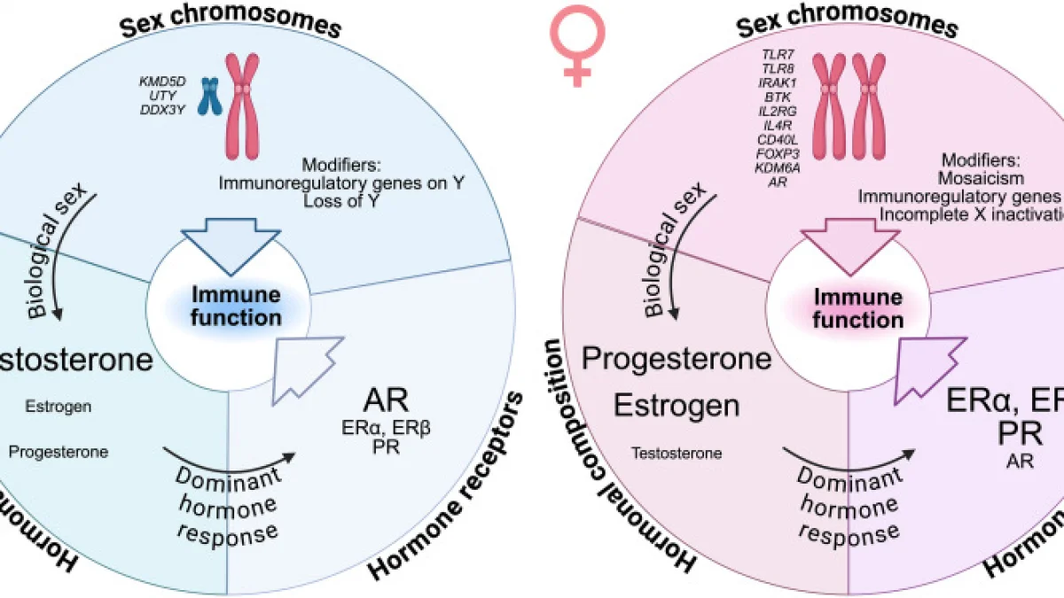
Biological sex exerts a profound influence on the baseline activation threshold, metabolic wiring, and functional plasticity of innate immune cells. These dimorphic responses stem from genetic, epigenetic, and hormonal regulatory networks:
1. X-Chromosome Inactivation Escape and Epigenetic Programming:
The X chromosome harbors a disproportionate number of genes critical for immune function. While one X chromosome is silenced during early female embryogenesis, approximately 15–20% of X-linked genes escape X-chromosome inactivation (XCI) in human immune cells. Endosomal *TLR7* and epigenetic modifiers such as lysine-specific demethylase 6A (*KDM6A* / *UTX*) and *KDM5C* frequently display biallelic expression in female myeloid cells. Elevated *TLR7* gene dosage lowers the threshold for endosomal nucleic acid sensing, leading to heightened Type I interferon production. Furthermore, KDM6A-mediated demethylation of histone H3 lysine 27 (H3K27me3) maintains an open chromatin conformation at pro-inflammatory gene promoters, maintaining female microglia in a chronically primed state.
2. Gonadal Steroid Receptor Modulation:
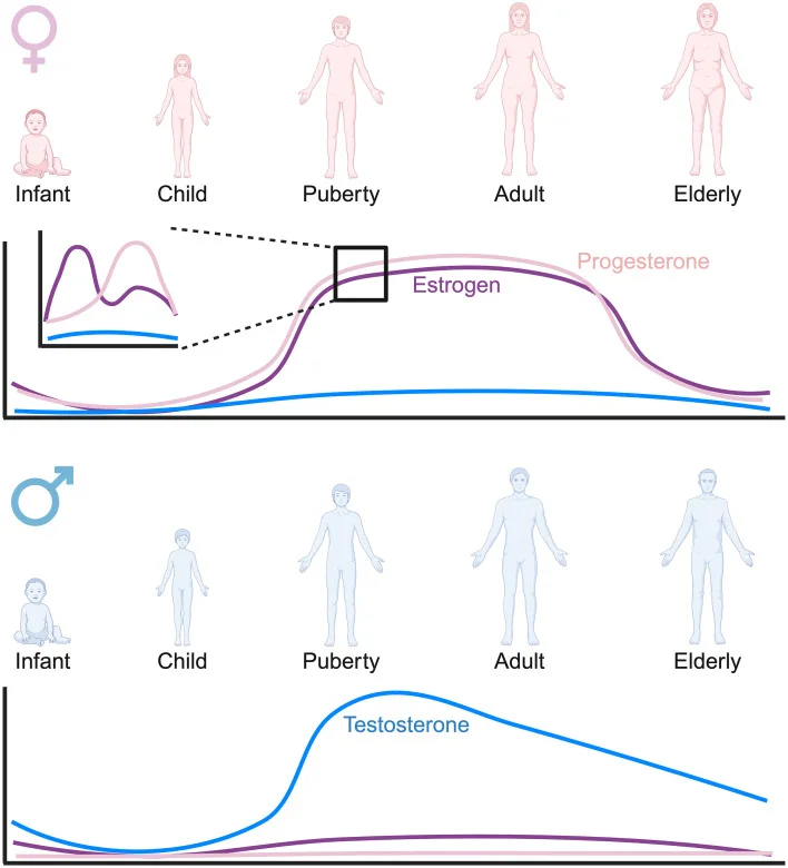
Sex hormones directly modulate myeloid transcription via intracellular signaling through estrogen receptors (ERα/ESR1, ERβ/ESR2), membrane G-protein coupled estrogen receptor 1 (GPER1), and the androgen receptor (AR). 17β-estradiol (E2) displays bimodal immunoregulation: low physiological concentrations promote classical pro-inflammatory (M1-like) microglial activation via ERα-mediated NF-κB transcription, whereas sustained high levels inhibit pro-inflammatory gene expression and upregulate neuroprotective pathways. In contrast, dihydrotestosterone (DHT) binding to AR recruits corepressors to NF-κB and AP-1 binding sites, suppressing NLRP3 expression and downstream IL-1β maturation, thereby conferring an immunosuppressive phenotype on male innate cells.
3. Translational Implications in Neurodegeneration and Immuno-Oncology:
In neurodegenerative CNS pathologies, female-biased microglial priming accelerates the transition toward Disease-Associated Microglia (DAM) states driven by TREM2-APOE signaling. While initial DAM activation facilitates phagocytic clearance of proteinaceous debris, chronic activation in females triggers hyper-inflammatory neurotoxicity, driving accelerated synaptic pruning and tau pathology in Alzheimer's disease models. Conversely, in oncological settings, this hyper-reactive baseline allows female tumor-associated macrophages (TAMs) to retain antigen presentation capacity and anti-tumor phagocytosis more effectively than male TAMs, which rapidly adopt immunosuppressive, pro-angiogenic phenotypes under AR and TGF-β control. Consequently, anti-cancer immunotherapies and anti-neuroinflammatory therapies exhibit marked sex-dependent efficacy profiles.
```
┌─────────────────────────────────────────┐
│ SEX-DIMORPHIC REGULATORY AXIS │
└────────────────────┬────────────────────┘
│
┌──────────────────────────────┴──────────────────────────────┐
▼ ▼
[ FEMALE AXIS ] [ MALE AXIS ]
• XCI Escape (TLR7, KDM6A) • Single X-chromosome Dosage
• ERα / GPER1 Activation • Androgen Receptor (AR) Activation
• Enhanced H3K27 Demethylation • Corepressor Recruitment (NF-κB/AP-1)
│ │
▼ ▼
• Lower TLR Activation Threshold • Elevated Activation Threshold
• Robust Type I IFN & mROS • Suppressed NLRP3 Inflammasome
• Primed Microglial/TAM State • Immunosuppressive/Pro-angiogenic TAMs
│ │
▼ ▼
• Accelerated Neuroinflammation • Diminished Anti-Tumor Immunity
• Superior Anti-Tumor Phagocytosis • Attenuated CNS Neurotoxic Pathology
```
Conclusions & Clinical Outlook
Sex-and-gender-based analysis (SGBA) represents a crucial frontier in precision biomedicine. The dimorphic molecular profiles of innate immune cells demonstrate that identical therapeutic interventions may yield disparate clinical outcomes between biological sexes. Future translational efforts must prioritize:
Integrating sex as a mandatory biological variable in preclinical drug development and clinical trial design will be indispensable for advancing targeted, high-efficacy therapeutics for neurodegenerative and oncological indications.
Key Insights
References
1. Klein, S. L., & Flanagan, K. L. (2016). Sex differences in immune responses. *Nature Reviews Immunology*, 16(10), 626–638. https://doi.org/10.1038/nri.2016.90
2. Guillot-Sestier, M. V., Doty, K. R., & Town, T. (2021). Innate immunity and neurodegeneration: Dynamic roles of microglia and systemic immune cells. *Journal of Neurochemistry*, 158(3), 601–619. https://doi.org/10.1111/jnc.15359
3. Spallanzani, R. G., Torres-Rojas, C., & Santambrogio, L. (2023). Dimorphic innate immunity in CNS and peripheral inflammatory disorders. *Frontiers in Immunology*, 14, 11864861. https://doi.org/10.3389/fimmu.2023.11864861